原文作者:Kevin Wright(兽医学博士 美国兽医从业人员执照持有者)
翻译:@诚熙L19
爬行天下ID:daibingrain
CANV金黄孢子菌
起因
在过去的文献里,我们把CANV作为导致许多疾病传染的根本原因而不是其他真菌。随着真菌培养与鉴定水平的发展,我们能更好的鉴别出是否CANV是爬虫患病的原因。CANV也是为数不多能完全符合柯赫氏法则的病原体之一。(即真菌从有临床症状的爬虫身上获取,然后在实验室中培养,再植入到其它爬虫体内,而另一只爬虫继续显示出同样的临床症状)(译者注:由于本人非兽医专业,此翻译未必专业,有兴趣者可自行百度或谷歌搜索“柯赫氏法则”)但最近的报告表明,从生病的爬虫身上又获取到了其它种类的Nannizziopsis(译者注:nannizziopsis为真菌界爪甲团囊菌科下一属,来源:维基百科)
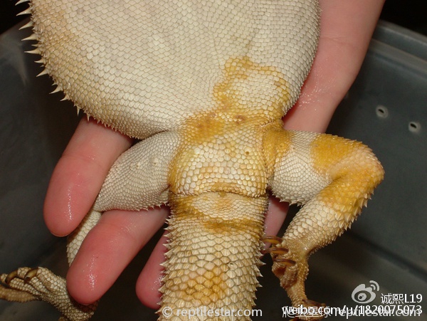
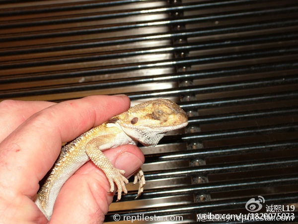
鬃狮蜥的CANV症状
处在危险中的物种
所有的爬虫都处在被感染的风险之中,特别是暴露在不洁的环境中,个体间拥挤或者紧张。鬃狮蜥,绿鬣蜥,高冠变色龙,王者蜥以及中国水龙等通常都是CANV的受害者。在一份已出版的吻突蝣蛇疾病与死亡报告中提到水栖爬行动物也有可能被传染CANV.
症状与预兆
CANV通常会表现为一小块黄色到棕色的小硬皮附着在一些鳞片的表面之上并随着时间推移越来越大。其他黄色到棕色的硬皮可能会随机在身体上任何地方扩散。极少数情况下,早期CANV患病的一个标志是一次不规则的蜕皮遗留下的有着粗糙表面的暗淡鳞片。在某些爬虫中可能会导致一场蜕皮风暴的到来。而这类爬虫也会几乎在蜕皮的开始就会变得暗淡与迟钝。随着病情的深入,这可能会导致爬虫内部感染,而最终导致食欲降低,失重甚至死亡。
如果你的饲养箱内有一只确诊的CANV爬虫患者,最好严密监视箱内其它个体是否有发病迹象
造成发病的饲养条件以及其它因素
CANV伴随着压力而来,通常见于拥挤而不洁的环境;某些群居的蜥蜴种类;被一起运输的个体;或被一起展示用于出售的个体。也有人认为在如此拥挤的环境下,脚趾甲和微小的咬伤伤口与物种内的相互传染相关,因为皮肤被损坏,进而真菌得以接种。环境卫生质量是很难与单一环境内的个体数量成正比,反而言之,处在如此环境下的个体就有了很大的可能性暴露给真菌。此外,拥挤导致的紧张也使得免疫反应得到抑制,所以单一个体也很可能无法对抗哪怕很低级别的真菌接种。CANV现在已经被发现可以存在于看起来健康的爬虫身上,也可以成为精心饲养下的爬虫的一种病因。
兽医可能推荐的诊断性测试
皮肤磨消法被推荐用来排除寄生虫(如螨虫)和检测是否为其它细菌性和真菌性感染。一份PCR诊断测试(译者注:PolymeraseChainReaction聚合酶链反应)可以发给专业实验室用来确认CANV的DNA是否存在。兽医也会建议采血以评估爬虫的健康状况以及排除一些严重的内在疾病。皮肤活体组织检查可帮助排除其它导致皮肤色素减退可能的原因。
安全操作与预防
隔离:如果你的饲养箱内有一只确诊的CANV爬虫患者,最好严密监视箱内其它个体是否有发病迹象。饲主应考虑将同笼的个体分开以降低从已明显感染个体与看起来健康个体之间交叉感染的风险。隔离的另一个原因是减少个体间的竞争状态。有时,爬虫间存在非常微妙的社会干预(译者注:通常我们遇到的个体成长不平衡,个体拒食或状态差的原因也很大与此有关)会导致紧张以及免疫力降低,进而更容易被感染。其余这些暴露在真菌环境影响下的个体也必须经过兽医检查与皮肤疾病评估从而排除可能潜在的CANV.
检疫:任何新进的带有皮肤破损的爬虫或在检疫期内发展为皮肤破损的个体,直到CANV作为可能存在的病因被彻底消除以后才可以被放到饲养环境。
正确的清洁与卫生:正确的清洁与卫生能把风险降到最低。如同其他传染病一样,一只正确投喂与悉心照料下的爬虫会有更好的能力抵御疾病的传染(例如:温度阶梯,合适的灯光,正确的湿度区间,躲避洞穴等)。避免购买拥挤环境下饲养的爬虫,特别是同一笼内已经存在有明显皮肤破损的个体。但很不幸的是,CANV普遍存在于人工圈养下的爬虫,一些爬虫会莫名其妙的患病。
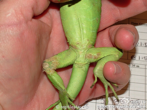
绿鬣蜥的CANV症状
兽医可能推荐的治疗方法
伏立康唑是一种已证明有可能有效的口服抗真菌药物,但是目前只有很少部分长期(超过24个月)的成功案例来证明。结合特比萘芬和伊曲康唑口服联合治疗,或口服结合两者其一,也未被证实长期有效。皮肤损伤应每日处以聚维酮碘一到两次。局部用药例如特比萘芬,咪康挫或伊曲康唑也未被证实长期有效。
预后
预后谨慎(译者注:是指预后的好坏,随病情的轻重、治疗是否得当及个体条件和环境因素的变化而有明显的不同。)大多数被感染的个体将会在未来12~18个月内死亡,也包括治疗过的。一部分已长时间幸存的个体也是处于长时间药物治疗,并且必须被认为是对其它爬虫的余生有着潜在的威胁。直到我们对治疗了解和研究的更多,否则CANV一定要被认为是一种威胁生命的疾病。
人畜共患的可能性
无
编者按:Wright博士于2013年9月6日意外死亡。时年51岁。当时他感刚刚完成发布于爬行动物杂志的一系列50种爬虫病例的论文。
译者的话:皮肤病应该是许多爬宠爱好者或者新手朋友们经常会遇到的问题。网上也有好多方法貌似对此能够有效,例如人用的治疗皮癣药物,温和消毒水甚至椰子油等方法。但是权威的研究似乎还没有对此过早下结论。危险随时可能会发生,我们能做到的就是积极的预防,以及一旦患病后耐心与细致的照料与治疗吧。
原文如下:
CANV in Reptiles
BY KEVIN WRIGHT, DVM, DABVP (RA)
Yellow fungus, Chrysosporium anamorph of Nannizziopsis vriesii, CANV
Causes
CANV is likely the underlying cause of many infections in older literature which attributed the disease to other fungi. Advances in fungal culture and identification helped distinguish CANV as a cause of disease in reptiles, one of the few pathogens that have fulfilled Koch’s postulates. (This means the fungus was recovered from a reptile showing clinical signs, was grown in a laboratory, was administered to another reptile, and that reptile went on to show the same clinical signs from which the fungus was recovered and demonstrated to be the same as the original isolate.) Recently there are reports of other species of Nannizziopsis being recovered from ill reptiles.
KEVIN WRIGHT
Bearded dragon with CANV.
Species At-Risk
All reptiles are at risk of infection, particularly when exposed to dirty conditions, crowding, and stress. Bearded dragons, green iguanas, veiled chameleons, Uromastyx, and green water dragons are commonly seen victims of CANV. Aquatic reptiles may also be affected as noted in one published report of illness and death of tentacled snakes from CANV.
Symptoms/signs
CANV often shows up as a small yellow to brown crust on the surface of a few scales that in time gets larger. Other yellow to brown crusts may appear elsewhere on the body in a random fashion. Occasionally, an early sign of CANV is an irregular shed that leaves behind dull scaled with a roughened appearance. In some reptiles it may result in a shedding storm where the reptile becomes opaque almost as soon as it has shed. As the disease progresses, it may become an internal infection and lead to poor appetite, weight loss, and death.
KEVIN WRIGHT
If you have a CANV positive reptile, any cage mate needs to be watched closely for signs of disease.
Predisposing captive conditions and/or other factors
CANV is associated with stress, often seen in reptiles kept under crowded unsanitary conditions. It’s often seen in those species of lizards which are raised in groups, shipped in groups, or displayed for sale in groups. It is also thought that toenails and minor bite wounds associated with intraspecific interactions under these crowded conditions results in damage to the skin and inoculation of the fungus. Sanitation may be compromised with high populations of lizards in the same cage which in turns increases the likelihood of a lizard being exposed to the fungus. Furthermore, crowding causes stress which results in suppressed immune response, so a lizard may not be able to fight even a low level inoculation of fungus. CANV has been recovered from seemingly healthy reptiles. It has also been a cause of illness in reptiles that were kept under good husbandry.
Diagnostic tests a veterinarian may recommend
A skin scrape may be recommended to rule out parasites like mites and to check for signs of other kinds of bacterial and fungal infections. A PCR test sent to a specialized lab may confirm the presence of CANV DNA. A veterinarian may suggest bloodwork to better assess your reptile’s health and rule out a serious internal illness. A skin biopsy may be needed to rule out other possible causes of skin discoloration.
Safe Practices / Prevention
Isolate: If you have a CANV positive reptile, any cage mate needs to be watched closely for signs of disease. You should consider separating the cage mates to reduce the risk of cross-infection from the obviously ill one to the seemingly healthy one. Another reason to separate them is to reduce competition between them. Sometimes there are subtle social interactions that create stress and immunosuppression, and make cage mates more susceptible to infections. Other exposed reptiles should have thorough veterinary examinations and any skin lesions should be assessed to rule out CANV as the underlying cause.
KEVIN WRIGHT
Green iguana with CANV.
Quarantine: Any reptile that arrives with a skin lesion or that develops a skin lesion while in quarantine should not be released until CANV is eliminated as the likely cause.
Proper hygiene and sanitation: Proper sanitation and hygiene are key risk reducers. As with other infectious diseases, a reptile is better able to resist an infection if it is fed the proper diet and is given excellent husbandry (i.e., a thermal gradient, appropriate lighting, the right range of humidity, hiding spots). Avoid purchasing reptiles kept under crowded conditions, particularly where there are obvious skin lesions on other specimens sharing the same container. Unfortunately, CANV is commonly present in the environments of captive reptiles and some reptiles inexplicably break with the disease.
Treatments a veterinarian would likely recommend
Voriconazole is an oral antifungal drug that has shown promise but there are few truly long-term (>24 mo) success stories at present. Oral combination treatments of terbinafine and itraconazole, or oral treatment with either alone, have not proved effective long-term. Skin lesions should be treated with povidone iodine once or twice daily. Topical treatment with antifungal drugs such as terbinafine, miconazole, or itraconazole, have not proven effective.
Prognosis
Guarded to poor. Most infected reptiles die within 12 to 18 months, even with treatment. The few that have survived long-term are often on long-term medical therapy and must be considered possible threats to other reptiles for the rest of their lives. Until more is known about treatment, CANV should be considered a life-threatening illness.
Zoonotic Potential
None.
Editor’s Note: Dr. Wright died unexpectedly on September 26, 2013. He was 51. He had just finished a series of 50 disease profiles for ReptilesMagazine.com when he passed.
